


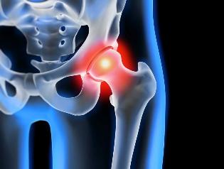
Osteoarthritis of the hip joint pathology degenerative that is characterized by destruction of hyaline cartilage. The disease develops gradually, associated with pain and reduced range of motion. In the absence of medical intervention in the initial phase of osteoarthritis several years later, there is atrophy of the thigh muscles. Wounds of the extremities are shortened, and the fusion of the joint space leads to partial or complete paralysis of the hip joint. Causes of the condition become previous trauma, curvature of the spine, systemic diseases of the musculoskeletal system.
Osteoarthritis usually is diagnosed in patients of middle age and the elderly. The diagnosis put on the basis of results of instrumental investigations — x-rays, MRI, CT, arthroscopy. The treatment of the disease 1 and 2 degrees of severity conservative. In the detection of ankylosis or ineffectiveness of drug therapy, surgical intervention (arthrodesis, arthroplasty).
The mechanism of the development of the disease
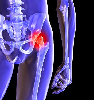
The hip joint consists of two bones — the iliac and femoral. The lower part of the Ilium presents her body, which participates in plexus with the femur, forming the upper part of the acetabulum. During the movement of the stationary glenoid fossa and the femoral head moves freely. This "hinge" device, the hip joint allows you to bend, stretch, rotate, contributes to the fun, bring the hips. The smooth sliding of the articulating structures provides soft, elastic, hyaline cartilage that covers the socket and the femoral head. Its main function is the redistribution of loads when driving, preventing rapid deterioration of the bone tissue.
Under the influence of external or internal factors alters trophic cartilage. You do not have a circulatory system — nutrients, tissues are supplied by the synovial fluid. In the case of osteoarthritis, which thickens and becomes slimy. The scarcity of nutrients, which causes a drying of the surface of hyaline cartilage. It is covered with cracks, which leads to a constant micro-trauma of the tissues in flexion or extension of the hip joint. The cartilages become thinner, lose their cushioning properties. To "adapt" to the increase of the pressure, the bones become deformed. And against the background of deterioration of metabolism in the tissues of the progress of the destructive-degenerative changes.
Causes and factors predisposing
Idiopathic or primary osteoarthritis develops without any reason. It is believed that the destruction of the cartilage occurs due to natural aging of the body, slowing down regenerative processes, the distribution of collagen and other compounds needed for the proper regeneration of the structures of the hip joint. Secondary osteoarthritis occurs on a background already present in the organism a condition of morbid. The most common causes of secondary disease are:
- previous injuries, damage of the tendon unit, muscle tears, its complete separation of the bone from the base of fractures, dislocations;
- developmental disorders of the joints, congenital dysplastic disorders;
- autoimmune disease — rheumatoid, reactive, psoriatic arthritis, systemic lupus erythematosus;
- nonspecific inflammatory diseases, such as suppurative arthritis;
- specific infections — gonorrhea, syphilis, brucellosis, anaplasmosis, trichomoniasis, tuberculosis, osteomyelitis, encephalitis;
- the interruption of the functioning of the endocrine system;
- pathology degenerative — osteochondropathy of the femoral head;
- hypermobility of the joints due to the production of "super traction" of collagen that trigger their excessive mobility, the weakness of ligaments.
Since the cause of development of osteoarthritis may be hemarthrosis (bleeding in the cavity of the hip joint), the precipitating factors include the violation of hematopoiesis. Requirements for the disease are excess weight, excess of exercise, sedentary lifestyle. For its development lead to a poor organisation of trainings, a deficiency in the diet products with a high content of minerals, fats and water soluble vitamins. Postoperative arthrosis occurs several years after the surgical intervention, especially if accompanied by the excision of a large volume of tissue. Osteoarthritis of the hip joint cannot be transferred by inheritance. But in the presence of certain congenital features (violation of metabolism, the structure of the skeleton), the probability that its development will be significantly increased.
Symptoms
The main symptoms of osteoarthritis of the hip joint pain when walking in the thigh, the knee. A person suffers from stiffness in the limbs, stiffness, especially in the morning. To stabilize the joint, the patient begins to limp, his gait changed. With time due to muscle atrophy and deformation of the joint of the limb is considerably reduced. Another characteristic feature of the pathology — limitation of hip abduction. For example, difficulties arise when one tries to sit on a stool, legs apart to the sides.
Even "launched" the ARTHRITIS can be healed at home! We need only remember once a day to smear it...
For osteoarthritis of the first degree is characterized by periodic pain that occurs after intensive physical loads. They are located in the area of articulation and disappear after a time of vacation.
The second degree of the arthrosis of the joints of the hip, the pain, the intensity increases. Discomfort is produced even at rest, are extended to the area of the groin, worse when lifting weights or increasing physical activity. To address hip pain, a person begins to barely noticeable limp. Marked limitation of motion of the joint, especially in abduction and rotation and internal hip.
Osteoarthritis of the third degree is characterised by constant severe pain. When the driving is difficult, therefore, at the time of walking of a person are forced to use a cane or crutches. Due to the weakness of abductor muscles of the hip is the offset of the pelvis bone on the plan front. To the extent that there was the shortening of the legs of the patient when the movement is tilted towards the limb affected. This causes a displacement of the center of gravity and increased stress on the joint. At this stage of the arthrosis develops a marked ankylosis of the joint.
| Degree | Radiographic signs |
| First | The changes are expressed not sharply. The joint cracks moderately, irregularly constricted, there is no destruction of the surface of the femur. In the interior or exterior of the edge of the acetabulum, there was a small bony growths |
| Second | The height of the joint space is reduced significantly due to their unequal sewing. Bone head of the femur is displaced upwards, deformed, enlarged, its edges become uneven. Osteophytes are formed on the surface of the inside and the outside of the edges of the glenoid fossa of |
| Third | There is a complete or partial fusion of the joint space. The head of the femur is much expanded. Multiple bone growths located on the surface of the acetabulum |
Diagnosis
At the time of establishing the diagnosis, the doctor considers that the clinical manifestations of the disease, anamnesis, external examination of the patient and instrumental examinations. The most informative radiography. With your help, the estimate of the state of the hip joint, which laid the foundations of its course, the degree of damage to cartilage tissues, and in some cases, the cause of development. If cervico-diaphyseal node is greater, and the acetabulum is flattened and beveled, then, with high probability it is possible to assume congenital dysplastic changes in the joints. On the disease of Perthes indicates a break in the form of the bone of the thigh. X-ray allows the identification and preparation of post-traumatic osteoarthritis, despite the lack of the former history of the disease of the injury. Also, use other diagnostic methods:
- CT helps detect the growth of the edges of the plates of bone formed osteophytes;
- An MRI is done to assess the condition of the connective tissue structure and the extent of their involvement in the pathological process.
If it is necessary, the inner surface of the joint is examined with the help of arthroscopic instruments. Differential diagnosis done to rule out osteoarthritis, lumbar-sacral or thoracic degenerative disk disease. The pain of osteoarthritis may masquerade as clinical manifestations of the syndrome is caused by pinching or inflammation of the nerve. To exclude neurogenic pathology general through a series of tests. Osteoarthritis of the hip joint necessarily differentialsa from the top of the bursitis of the hip, ankylosing spondylitis, reactive arthritis. To exclude autoimmune pathologies are conducted biochemical studies of blood and synovial fluid.
Surgery
With the ineffectiveness of therapy conservative Il diagnosis complicated pathology of the surgery performed. To restore the cartilage of joints damaged by arthritis, no surgeries impossible, but with the right approach of treatment, the compliance with all rules, keep proper life style, gymnastics medical, courses of massage, the intake of vitamins and proper nutrition can stop the process of destruction, and the destruction of cartilage and joints of the hip.